The aims of this research were to understand whether pericytes, a cell located on the smallest blood vessels of the brain (capillaries), are implicated in how the brain responds to an ischaemic stroke, and whether targeting pericytes could improve stroke outcomes.
Aim 1: To determine how depletion of pericytes alters injury after stroke
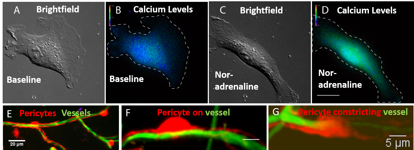
To determine how pericytes may influence stroke outcomes, a method was developed that could deplete pericytes. A number of cell culture studies were carried out, and identified two molecules that killed pericytes by blocking a receptor on their surface (platelet derived growth factor receptor beta (PDGFRβ)). Unfortunately, there was no clear reduction in pericyte number or coverage of blood vessels in the brain (Fig 1E) when these drugs were administered in mice. An alternative strategy was developed to deplete pericytes using a genetic mouse model which enabled a reduction in pericyte numbers by 90% in the brain. A novel method to monitor blood flow in the brain was also developed, which helps determine how pericyte depletion affects blood flow.
Aim 2: To identify mechanisms that mediate pericyte contraction and relaxation to alter blood flow
The aim was to determine how pericytes regulate blood flow by contracting or relaxing blood vessels. A novel imaging technique that enabled us to observe pericytes contracting and relaxing in real-time was developed. It was identified that calcium was a master regulator of pericyte contraction to a number of vasoactive mediators (Fig 1A-D). Numerous molecules were tested to either prevent pericyte contraction or promote pericyte relaxation. It was discovered that a common immunosuppressant drug ordinarily used for transplantation, could block pericyte contraction.
Aim 3: To block pericyte contraction or promote pericyte relaxation to protect the brain after stroke
The most promising molecules from the cell culture work (Aim 2) were taken into animal studies, to look at the efficacy of using a drug that can target pericytes on stroke outcomes. The immunosuppressant drug was a clear candidate that could target pericyte contraction. Experiments were conducted (and a meta-analysis) showing that it can reduce lesion volume in a mouse model of stroke, and this appears to be due to an improvement in blood flow. Experiments are underway in mice whereby the immunosuppressant is administered during a stroke to determine whether it can prevent pericyte contraction following stroke (Fig 1F, G), giving rise to their protective effects on the brain. These studies could inform whether immunosuppressant drugs that act on specific mechanisms within pericytes would be a useful repurposed drug for the treatment of ischaemic stroke.
Conclusion and future directions. Important research was carried out to establish a method to deplete pericytes in the mouse brain, uncover a novel drug that can block pericyte contraction, and that can protect the brain following stroke. These initial data have enabled A/Prof Sutherland to secure further funding from NHMRC for this project, and fully establish pericytes as a novel cellular target for the treatment of ischaemic stroke.
Figure: Brightfield image of a pericyte in cell culture (A) and its baseline calcium levels (B). Following exposure to noradrenaline, pericytes contracted (C) and had increased calcium levels (D). Mice that have fluorescent (red) pericytes can be visualised on blood vessels (green) to determine pericyte number and coverage of vessels (E). Two morphologies of pericytes were observed, (F) pericytes on a vessel, and (G) pericytes wrapping around a vessel.