Optical coherence tomography (OCT) is an imaging technique capable of rapid non-invasive acquisition of high-resolution images of the eye, providing an “optical biopsy” of the ocular tissues, on a micron scale. OCT imaging has transformed the ophthalmic field and is a standard imaging modality routinely used to assess the health of the eye, as it provides fundamental information for clinical decision making.
Visual image inspection by an eye practitioner provides useful information, but structural (thickness) analysis represents the key quantitative information extracted from these medical images. This thickness information extracted from OCT images is a fundamental marker used in the evaluation of ocular health, disease detection/progression and treatment outcomes. Commercial OCT instrument software does not accurately measure all of the tissue layers (such as the vascular choroidal layer) and commonly fails to quantify retinal thickness, especially in the presence of pathology. This means that time-consuming manual segmentation of OCT images is often required.
In this project, a range of image segmentation tools (defining the position of tissue layers) for OCT images of the posterior part of the eye, were developed. These tools are based on deep learning techniques, a branch of artificial intelligence. Deep learning methods take advantage of previously segmented OCT images to learn patterns in the data through an initial “training phase”. During this phase, hundreds of images are presented to the deep neural network to “learn” the patterns. After this training phase, new “unseen” OCT images can be passed through the network to obtain an automatic segmentation of the OCT image. This project has developed novel neural network architectures for medical image analysis.
The different tools that were developed during the project have accomplished a number of objectives:
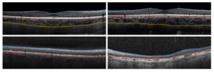
- Novel choroid segmentation techniques: the proposed method allows accurate extraction of layer information (the vascular choroidal layer) that are not possible with current commercial instruments.
- Pathology segmentation techniques: a method was proposed to accurately and automatically segment the retinal tissue for OCT images from patients with Stargardt disease (an inherited macular degeneration). This can have major impact on data analysis and clinical decision making.
- Synthetic OCT data generation: given the importance of labelled data in deep learning methods, a novel method for synthetic OCT data generation was developed. This synthetic data can be used to train the network and decreases the need for large data training sets.
These tools are open source, to ensure other teams can benefit from this research and the clinical and research impact of these tools can be easily disseminated. The developed method can be applied to other image modalities or OCT disease applications.
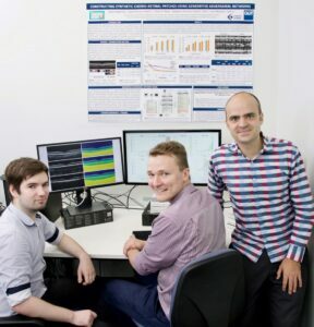
Picture below: Principal investigator (Dr David Alonso-Caneiro [right]) with two of the research engineers involved in the project (Jared Hamwood [left] and Jason Kugelman [centre]).
Picture below: Example OCT images with the automatic retina and choroidal boundary segmentation in healthy image (top plots) and images from patients with Stargardt disease (bottom plots). The retinal layer is located between the blue and red boundaries, while the choroidal layer is located between the red and yellow (not detected in Stargardt disease patients).