Funding from the Rebecca L Cooper foundation supported several papers and conference presentations. Highlights include:
Large-scale brain modes reorganize between infant sleep states and carry prognostic information for preterms: This paper showed that the brain activity in sleeping infants born preterm differs from that of infants born at term. In term infants there is a large reorganisation of brain activity between the two sleep states of “active” and “quiet” sleep (infant versions of adult rapid eye-movement sleep and deep sleep). In preterm infants measured at term age, this reorganisation is weaker. Using methods from physics Dr Roberts found a biophysical mechanism for how these differences arise. Moreover, it was found that the differences relate to neurodevelopmental outcome at age 2 years. This work thus provides new understanding of infant brain dynamics and offers potential new prognostic markers of infant brain health.
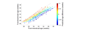
Automated cot-side tracking of functional brain age in preterm infants: In this paper, Dr Roberts’s group showed proof-of-principle for “growth charts” for preterm infant brain function. It is standard to track a baby’s weight, height, and head circumference to verify that the baby is developing “correct for age”. But there are no tools for verifying that brain function is similarly tracking along a healthy path. Using machine learning, the team developed and validated a method for checking that a preterm baby’s brain activity is “correct for age”. Results also showed that being behind early on can predict adverse neurodevelopmental outcomes at age 2 years. This work shows that a new diagnostic tool (“functional brain age”) can be used to accurately track infant brain development, potentially enabling better and earlier targeting of treatments for babies most at need.
Reliability and accuracy of EEG interpretation for estimating age in preterm infants: In a follow-up paper, it was shown that the research team’s functional brain age algorithm can outperform human expert raters. Assessing that brain activity is correct for age is a common task for clinician experts who interpret traces of brain signals. But they do it by eye in a time-consuming process, and inevitably there are inconsistencies between different raters. The automated algorithm gives more precise estimates than human experts, and with less bias. This work shows that brain function growth charts have the potential to augment or even replace expensive visual interpretation.
Automated detection of artefacts in neonatal EEG with residual neural networks: This paper addresses important technical hurdles toward getting methods such as the above into the neonatal intensive care unit. Real-world brain recordings are often contaminated by artefacts from outside the brain (e.g. interference from other hospital machines, heartbeats, muscle movements, etc.). To use algorithms at the cot-side requires handling of these artefacts. This study used machine learning to develop an automated artefact detector. This new tool not only detects artefacts but categorises them too, opening the possibility of identifying the source of the contamination. This algorithm will be an important piece of a final real-world implementation of automated diagnostics in the clinic.
Looking ahead, Dr Roberts has recently partnered with a brain monitoring device company to take these algorithms to the next step toward translation. Real-time implementations of these algorithms are being developed for use with clinical monitors, and will be trialled at Royal Brisbane and Women’s Hospital.