Epithelial mesenchymal transition (EMT) can contribute to small airway fibrotic pathology in smokers and patients with COPD. Myofibroblasts are highly active pro-fibrotic cells that secrete excessive and altered extracellular matrix (ECM).
In the study published by Dr Sohal in the European Respiratory Journal Open Research, small airway myofibroblast presence was related with airway remodelling, physiology and EMT activity in smokers and COPD patients.
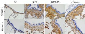
Lung resections from non-smoker controls, normal lung function smokers and COPD current and ex-smokers were stained with anti-human α-smooth muscle actin (SMA), collagen 1 and fibronectin. αSMA+ cells were computed in reticular basement membrane (Rbm), lamina propria and adventitia and presented per mm of Rbm and mm2 of lamina propria. Collagen-1 and fibronectin are presented as a percentage change from normal. All analyses including airway thickness were measured using Image-pro-plus 7.0.
An increase in subepithelial lamina propria and adventitia thickness in all pathological groups compared to non-smoker controls was found. Increases in αSMA+ myofibroblasts were observed in subepithelial Rbm, lamina propria and adventitia in both the smoker and COPD groups compared to non-smoker controls. Furthermore, the increase in the myofibroblast population in the lamina propria was strongly associated with decrease in lung function, lamina propria thickening, increase in ECM protein deposition, and finally EMT activity in epithelial cells.
This is the first systematic characterisation of small airway myofibroblasts in COPD based on their localisation, with statistically significant correlations between them and other pan-airway structural, lung function and ECM protein changes.
Dr Sohal and his team suggest that EMT may be involved in such changes. The data collected would support the logic and need for treatment as early as possible and identifies possible new pathophysiological targets for therapy. In-vitro data identified the effect of M2 macrophage chemokine/cytokine treatment on gene expression of mesenchymal markers such as N-cadherin and Vimentin and epithelial junctional protein E-cadherin in primary small airway epithelial cells. Addition of M2 macrophage secretory protein CCL22 was found to markedly enhance N-cadherin genes expression over two-fold compared to TGFβ1 alone and close to nine-fold over control. IL-4 also showed similar increases albeit lesser than CCL22. E-cadherin gene was found to be supressed across all treatment in comparison to cell controls.
Similar changes in type 2 pneumocytes as well in response to these stimulants plus other environmental factors such as diesel exhaust particles were observed. Data suggested that both small airways epithelial cells and type 2 pneumocytes could lead to fibrosis through EMT under the influence of pro-fibrotic M2 macrophage environment. Epithelial-hybrid mesenchymal spectrum in COPD was also measured using transcriptomic-based quantification. The findings were compared to patients with idiopathic pulmonary fibrosis and normal controls, which indicated a significant mesenchymal cell load in COPD.
Lungs of smokers and COPD patients are highly susceptible to infections which could lead to fibrotic changes through EMT. Given the fact this project was going through the COVID environment presented the opportunity to look if these patients are more susceptible to SARS-CoV-2 leading to post COVID interstitial fibrosis. SARS-CoV-2 attachment receptor ACE2 increase on alveolar macrophages was observed along with endocytic vacuoles such as EEA1, RAB7, Cathepsin-L and LAMP1. Increased type 2 pneumocytes suggested that these patients are vulnerable to developing post-COVID-19 interstitial pulmonary fibrosis or fibrosis in general. There could be a silently developing interstitial pathology in smokers and patients with COPD. Active TGF-β1 and pSmad2/3 transcription factors promoting EMT activity was found in the same tissue from smokers & COPD patients. Hence, EMT could be central to these fibrotic changes driven by infections and inflammation.
Image: surgically resected lung tissue (taken at open chest surgeries) stained for markers of EMT from normal lung function smokers (NLFS), COPD current smokers (COPD-CS) and COPD ex-smokers (COPD-ES) compared to normal controls (NC).