
Vulvodynia causes severe pain, sexual dysfunction, shame and distress for millions of young women and girls. For 30 years it has been known that the most common form of vulvodynia, neuroproliferative vestibulodynia, involves changes in vaginal nerves. However, limited knowledge of these nerves has led to there being no effective medical treatment for neuroproliferative vestibulodynia.
This study has successfully shown that mild inflammatory stimulus at the vaginal entrance causes migration of specific immune cells to the area along with growth of vaginal nerves and increased pain sensitivity. In addition, pain-associated signalling also increases in the spinal cord.
This project also showed that a drug commonly used to treat bone weakness is very effective in preventing pain sensitisation in this model. This drug also reduces the abundance of the immune cells (macrophages), and normalises nerve to nerve signalling in the spinal cord. This provides evidence that signalling between macrophages and nerves in the vagina may contribute to the key pathological processes involved in the most common form of vulvodynia. Molecules involved in macrophage-sensory nerve signalling pathways present useful molecular targets to better understand and treat vestibulodynia.
Dr Barry’s ongoing research investigates these pathways in her model compared to contemporary clinical studies.
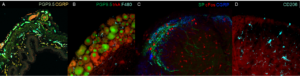
Image description:
These microscope image show sections of mouse tissue labelled for nerves responsible for sensation. Specific signalling molecules that regulate pain sensitivity are also labelled. The nerves we study have cell bodies in dorsal root ganglia, peripheral endings in the vagina and central endings in the lumbosacral spinal cord. Changes in any of these regions can contribute to chronic pain.
A. Cross-section of vagina showing abundant nerve endings labelled by protein gene product (PGP9.5, pale green). Inflammation causes these nerve endings to proliferate. Many of these nerves contain calcitonin gene-related peptide (CGRP, yellow), a small protein that causes blood vessels to dilate and pain sensitivity to increase.
B. Mouse dorsal root ganglia showing the round cell bodies sensory nerves labelled by PGP9.5 (green). Nerves containing the signalling molecule trkA (red) respond to nerve growth factor. Macrophages (cyan) are immune cells that can be seen located between nerve cell bodies and may contribute to pain sensitivity.
C. Spinal cord region where sensory nerves from the vagina signal to nerves of the central nervous system. cFos (red) shows activated spinal cord nerves. Central projections of specific sensory nerves from the periphery are shown by labelling for Substance P (SP, green) which contributes to pain signalling and CGRP (red), which increases the response of spinal cord neurons to substance P.
D. The spinal cord also contains immune cells that contribute to regulation of pain sensitivity. This micrograph shows spinal cord microglia labelling by the cell-surface marker CD206